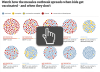
You and Your Health
the molecules that keep us alive
Healthy molecules help build a healthy body. In order for our cells to function properly, all of their molecules need to be performing their individual jobs. Problems occur, for instance, when a key vitamin is missing from our diet or a protein is compromised by a faulty gene. Atomic structures have revealed how healthy molecules function and how errors in this function can cause disease.
Molecule of the Month Articles (59)
| ABO Blood Type Glycosyltransferases
ABO blood types are determined by an enzyme that attaches sugars to proteins |
|
 |
Acetylcholinesterase
Acetylcholinesterase stops the signal between a nerve cell and a muscle cell |
 |
Alcohol Dehydrogenase
Alcohol dehydrogenase detoxifies the ethanol we drink |
 |
Amyloid-beta Precursor Protein
Cell-clogging amyloids form when proteins improperly aggregate |
 |
Amyloids
Alzheimer's disease and prion diseases are linked to unnatural aggregation of proteins into amyloid fibrils. |
 |
Anabolic Steroids
Anabolic steroids like testosterone are among the most common performance enhancing drugs |
 |
Angiotensin and Blood Pressure
Many medications for controlling high blood pressure inhibit the action of the peptide hormone angiotensin. |
 |
Apoptosomes
Apoptosomes make life or death decisions in cells |
 |
Beta-secretase
Beta-secretase trims proteins in the cell and plays an important role in Alzheimer's disease |
 |
Calcium Pump
Atomic structures have captured the calcium pump in action |
 |
Carbonic Anhydrase
Carbonic anhydrase solubilizes carbon dioxide gas so we can breathe it out |
 |
Carotenoid Oxygenase
Light-sensing retinal molecules are built from colorful carotenoids in our diet |
 |
Caspases
Caspases disassemble proteins during the process of programmed cell death |
 |
Catalase
Catalase protects us from dangerous reactive oxidizing molecules |
 |
CFTR and Cystic Fibrosis
Cystic fibrosis is currently treated using drugs that enhance the function of mutated CFTR |
 |
Circadian Clock Proteins
Circadian clock proteins measure time in our cells |
 |
Collagen
Sturdy fibers of collagen give structure to our bodies |
 |
Crystallins
A concentrated solution of crystallins refracts light in our eye lens |
 |
Cytochrome p450
Cytochrome p450 detoxifies and solubilizes drugs and poisons by modifying them with oxygen |
 |
DNA Methyltransferases
Cells add methyl groups to their DNA to encode additional epigenetic information |
 |
Estrogen Receptor
Estrogen binds to receptors in the nucleus and affects key genes in development |
 |
Fetal Hemoglobin
Fetal hemoglobin allows a growing fetus to receive oxygen from their mother. |
 |
Fibrin
Rod-shaped fibrin molecules link together to form blood clots |
 |
Glucansucrase
Bacteria adhere to our teeth by building sticky sugar chains |
 |
Growth Hormone
Growth hormone brings together two copies of its cellular receptor |
 |
Hemoglobin
Hemoglobin uses a change in shape to increase the efficiency of oxygen transport |
 |
Hyaluronidases
Long carbohydrate chains are used to make our bodies flexible and resilient. |
 |
Hypoxanthine-guanine phosphoribosyltransferase (HGPRT)
Cells salvage and recycle their obsolete DNA and RNA |
 |
Hypoxia-Inducible Factors
HIF-α is a molecular switch that responds to changing oxygen levels. |
 |
Insulin
The hormone insulin helps control the level of glucose in the blood |
 |
Leptin
Problems with the appetite-controlling hormone leptin can lead to obesity |
 |
Lysozyme
Lysozyme attacks the cell walls of bacteria |
 |
Major Histocompatibility Complex
MHC displays peptides on the surfaces of cells, allowing the immune system to sense the infection inside |
 |
Neurotrophins
Neurotrophins guide the development of the nervous system |
 |
Nicotine, Cancer, and Addiction
Nicotine causes addiction by interacting with receptors in the brain |
 |
Opioid Receptors
Morphine and other opioid drugs bind to receptors in the nervous system, controlling pain |
 |
Oxidosqualene Cyclase
Oxidosqualine cyclase forms the unusual fused rings of cholesterol molecules |
 |
Pepsin
Pepsin digests proteins in strong stomach acid |
 |
Phenylalanine Hydroxylase
An unusual cofactor is used in the synthesis of aromatic amino acids |
 |
Phospholipase A2
Phospholipase A2 breaks membrane lipids, forming molecules that contribute to inflammation and pain signaling. |
 |
Piezo1 Mechanosensitive Channel
Mechanosensitive ion channels give our cells a sense of touch. |
 |
Prions
Mad cow disease is caused by prion proteins that misfold and aggregate |
 |
Proton-Gated Urea Channel
A channel that passes urea allows ulcer-producing bacteria to live in the stomach |
 |
S-Nitrosylated Hemoglobin
Nitric oxide is attached to a conserved cysteine in hemoglobin and then released to control the flow of blood. |
 |
SARS-CoV-2 Nucleocapsid and Home Tests
Home test kits for SARS-CoV-2 test for the presence of nucleocapsid, the protein that packages the viral genome in infectious virions. |
 |
Selenocysteine Synthase
Selenium is used in place of sulfur to build proteins for special tasks |
 |
Serotonin Receptor
Serotonin receptors control mood, emotion, and many other behaviors, and are targets for many important drugs |
 |
Serpins
Serpins are traps that capture dangerous proteases |
 |
Serum Albumin
Serum albumin delivers fatty acid molecules through the bloodstream |
 |
Sirtuins
Sirtuin activation is being explored as a way to slow aging. |
 |
Superoxide Dismutase
Superoxide dismutase protects us from dangerously reactive forms of oxygen |
 |
T-Cell Receptor
Lymphocytes use T-cell receptors to patrol the body for foreign molecules |
 |
Tetrahydrobiopterin Biosynthesis
Tetrahydrobiopterin plays an essential role in the production of aromatic amino acids, neurotransmitters and nitric oxide. |
| Thrombin
Thrombin activates the molecule that forms blood clots |
|
 |
Tissue Factor
Tissue factor senses damage to the body and triggers formation of a blood clot |
| Tissue Transglutaminase and Celiac Disease
Tissue transglutaminase staples proteins together by forming a chemical crosslink. |
|
 |
Vitamin D Receptor
Vitamin D helps regulate the use of calcium throughout the body |
 |
Vitamins
Vitamins are essential molecular tools that are obtained through a healthy diet. |
 |
Xanthine Oxidoreductase
Xanthine oxidoreductase helps break down obsolete purine nucleotides |
Learning Resources (10)
 |
Insulin
Paper Model
Learn about insulin, a peptide hormone that plays a critical role in our ability to use glucose from the food that we eat
|
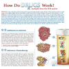 |
How do Drugs Work?
Flyer
PDB structures are used to discuss antibiotics and antivirals, chemotherapy, drug metabolism, drugs of signaling proteins, and lifestyle drugs.
|
 |
How Do Drugs Work?
Poster
PDB structures are used to discuss antibiotics and antivirals, chemotherapy, drug metabolism, drugs of signaling proteins, and lifestyle drugs.
|
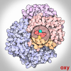 |
Oxygen Binding in Hemoglobin
GIF
Hemoglobin uses a change in shape to increase the efficiency of oxygen transport.
|
 |
Opioids and Pain Signaling
Video
Pain is one of the most trying experiences of life. On the cellular level it is communicated via special neuronal pathways. On the molecular level, however, pain is communicated like any other sensation, via a set of electrical and chemical signals facilitated by complex molecular machinery. These signals can be modulated by opioids, causing us to feel less pain, or no pain at all. Learn how opioids activate the G-proteins which in turn interact with other proteins to edit the pain signal.
|
| Calcium Pump
Video
The calcium pump moves ions across cell membranes allowing the synchronized contraction of muscle cells.
|
|
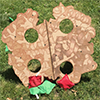 |
Hemoglobin Bean Bag Toss
Other Resource
|
| Passive Immunization with Convalescent Antibodies
Article
Purified antibodies may be used to treat infection by coronavirus.
|
|
| The Search for Drugs to Fight COVID-19
Article
Medical researchers are testing previously-discovered antiviral drugs for effectiveness against SARS-CoV-2.
|
|
| Dexamethasone and Cytokine Storms
Article
Preventing too much of a good thing during SARS-CoV-2 infection
|
Curriculum Resources (14)


Structural Biology Highlights (6)
Global Health (10)
| Diabetes Mellitus - What is Diabetes?
Diabetes mellitus is a metabolic disorder that affect many lives globally.
|
|
| Diabetes Mellitus - Causes of Diabetes
Diabetes can be caused by the absense or improper function of insulin.
|
|
| Diabetes Mellitus - Types of Diabetes
Diabetes is classified into various types based on its causes.
|
|
| Diabetes Mellitus - Statistics
Diabetes is a growing global health concern.
|
|
| Diabetes Mellitus - Timeline
This timeline lists key events in the discovery of Diabetes and its main treatment options.
|
|
| Diabetes Mellitus - Symptoms | |
| Diabetes Mellitus - Diagnosis
Diabetes is diagnosed by measuring blood or plasma glucose levels and/or levels of hemoglobin glycation
|
|
| Diabetes Mellitus - Complications
Uncontrolled diabetes can lead to many acute and chronic complications
|
|
| Diabetes Mellitus - Non-Pharmacological
Lifestyle changes, including improvement in diet and increasing exercise, can help manage and prevent progression of diabetes.
|
|
| Diabetes Mellitus - Pharmacological
A number of pharmacological options are available to treat diabetes.
|
Geis Digital Archive (15)
 |
Lysozyme (512)
Geis illustrates the structure of lysozyme, which was first revealed by X-ray crystallography in 1965 (Blake et al., 1965). The structure of lysozyme was the first to be determined via this method. Geis carefully highlights the interaction between lysozyme and the substrate. This particular illustration appeared on the cover of Scientific American Volume 215, Issue 5 (Phillips, 1966). |
 |
Hemoglobin S
Geis illustrates the hemoglobin s molecule as four nearly symmetrically arranged subunits with a mutation present in the beta chain.
|
 |
Oxyhemoglobin
Geis illustrates the hemoglobin molecule in its oxygenated state.
|
 |
Hemoglobin
Geis illustrates the hemoglobin molecule as four symmetrically arranged myoglobins. Since it is responsible to the transport of oxygen, it can change from an oxygen-binding configuration to an oxygen-releasing configuration in response to the demand for oxygen. |
 |
Hemoglobin (1000)
Geis uses four colors to depict the hemoglobin tetramer structure. The heme groups are shown as white rectangular prisms. A red sphere is in the middle of each of these, representing an iron ion that allows capture of oxygen molecules. Hemoglobin is essential in transporting oxygen in vertebrates. |
 |
DPG-Hemoglobin Complex
Geis illustrates the complex of DPG (2,3-diphosphoglycerate) with hemoglobin as a co-crystal. The charged amino acid residues stabilizing the complex with DPG are drawn in blue. |
 |
Deoxyhemoglobin
Geis illustrates deoxyhemoglobin in blue, a color associated with the absence of the oxygen molecule. Hemoglobin undergoes a conformational change in the absence of oxygen to produce this conformer. In the center is a red area showing the footprint of the space between the beta chains in the oxygenated state. The beta chains move apart in the deoxygenated state.
|
 |
Intermolecular Contacts in Hemoglobin S
Geis paints the intermolecular contacts in hemoglobin S. The lateral contacts are formed by the interactions between residues 66, 73, 80, 83 and 87 of one beta subunit and a mutant beta 6 valine from an adjacent molecule. The axial contacts are made by interactions along the molecule and include residues 16,17,19,22 and 121. Residues 95,47 and 75 interact through contacts between twisted filaments of the molecule.
|
 |
Cytochrome c
Geis illustrates cytochrome C, one of the proteins responsible for energy transfer in the electron-transport chain. He highlighted the central heme group in the molecule. Cytochrome C is essential to energy production in the body, and this particular function is highlighted by Geis' choice of framing the central heme as a source of light. |
 |
Deoxyribonucleic Acid (DNA)
Geis illustrates three possible forms of deoxyribonucleic acid (DNA). He highlights the differences between each structure by displaying them in a side-by-side manner. |
 |
B-DNA
Geis illustrates B-DNA in blue looking from above, through the double helix. The two bases on top are highlighted in white to distinguish one individual section of the layered scene. |
 |
A-DNA
Geis uses a thin ball and stick representation of a section of A-DNA, the more compact conformation of DNA less often seen in biological systems. He draws it from a perspective looking down into the double helix, showing the increase in diameter of the middle of the helix from the B form DNA.
|
 |
Carboxypeptidase A
Geis depicts the structure of carboxypeptidase A, highlighting the central twisted beta sheet of the enzyme. The enzyme itself cuts polypeptide chains from the carboxyl terminal end. Included in the painting is an orange dot representing a zinc atom at the active site.
|
 |
Collagen
Geis paints a triple twist pattern in his depiction of collagen. He portrays the chains in a thick, rope-like manner, which reflects the function of the collagen protein in strengthening tendons and muscles in animals. |
 |
Lac Repressor
In this acrylic painting of the lac repressor, Geis characterizes this tetramer representation as "four angry reindeer." Geis' painting depicts the lac repressor with both the tetramerization domain (on the bottom) and the headpieces ("antlers" of the "reindeer"). A complete crystal structure of this tetramer has not actually been determined yet. |
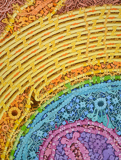
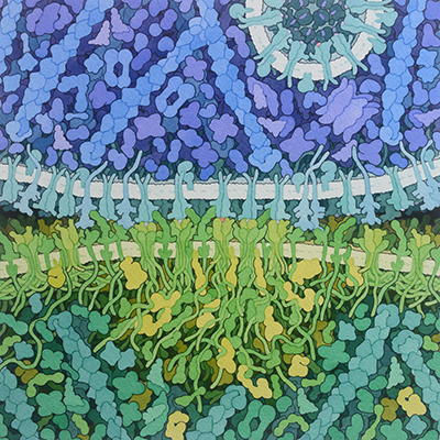
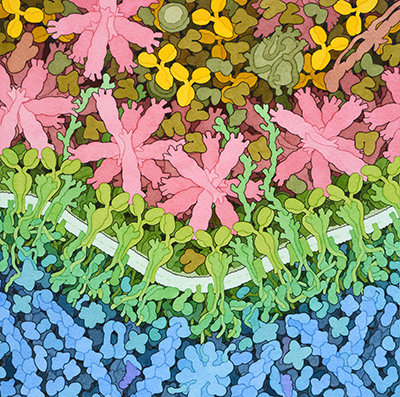
Goodsell Molecular Landscapes (9)
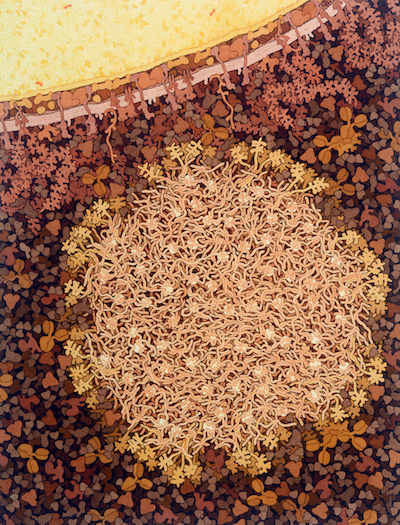 |
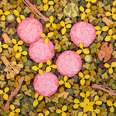
Casein Micelle and Fat Globule in Milk
Illustration of a casein micelle and fat globule from milk
|
 |
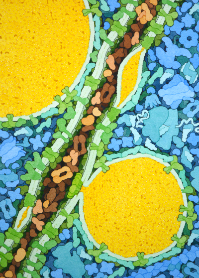
Myelin
Myelin forms an insulating sheath around nerve axons
|
 |
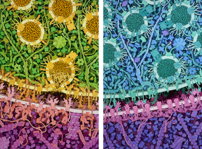
Immunological Synapse
Antigen-presenting cells stimulate T-cells with small viral peptides.
|
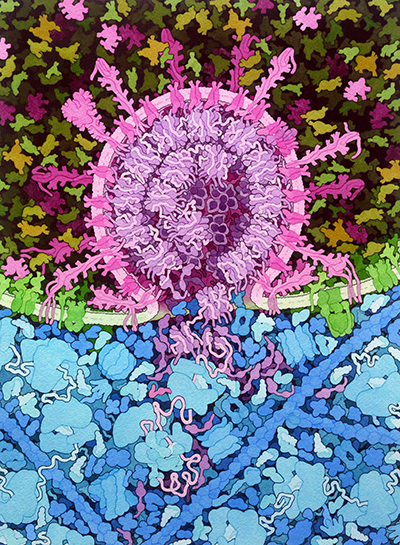 |
SARS-CoV-2 Fusion
SARS-CoV-2 fuses with endosomal membranes, releasing the viral RNA genome into the cell.
|
 |
Influenza Vaccine
Influenza vaccine causes the production of antibodies that fight infection by the virus
|
 |
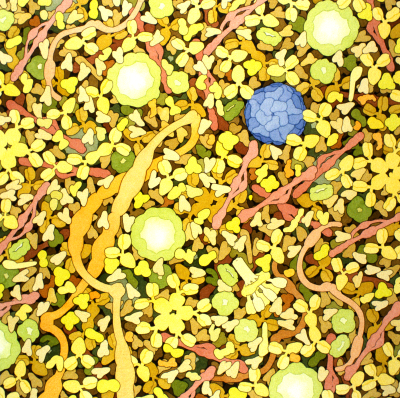
Lipid Droplets
Lipids such as fats and cholesterol are stored in large droplets inside cells.
|
 |
Poliovirus Neutralization
Polio vaccine causes the immune system to create antibodies that neutralize poliovirus
|
 |
Excitatory and Inhibitory Synapses
Excitatory and Inhibitory Synapses (2018) by David S. Goodsell
|
 |
Biosites: Blood Plasma
Biosites: Blood Plasma (2005) by David S. Goodsell.
|
