
Style
Color
Spin
Guide to Understanding PDB Data
Guided reference for methods of obtaining, exploration, and interpretation of PDB entries
Recorded lectures and related resources
A Deep Dive into Computed Structure Model Exploration at RCSB.org


Images used with permission from the Howard Hughes Medical Institute (www.hhmi.org). All rights reserved.
Induced Lac Repressor
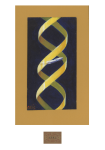
Z-DNA
TATA-Binding Protein (TBP)
Myoglobin Fold
Collagen
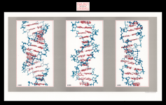
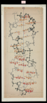
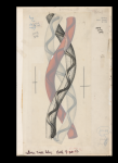
Deoxyribonucleic Acid (DNA)
DNA
Transfer Ribonucleic Acid (tRNA)
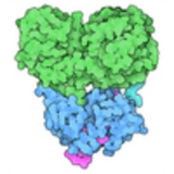
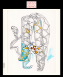
Aspartate Transcarbamoylase (ATCase)
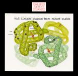
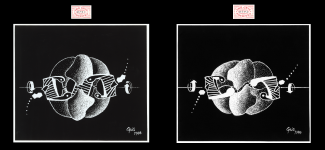
Intermolecular Contacts in Hemoglobin S
Lysozyme (488)
Cytochrome c (unbound)
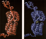
Cytochrome c
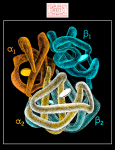
Lac Repressor
Hemoglobin S
Myoglobin
Oxyhemoglobin
Deoxyhemoglobin
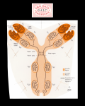
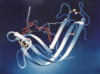
Immunoglobulin G (IgG)
A-DNA
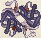
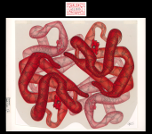
Hemoglobin
Myohemerythrin
Carboxypeptidase A
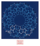
Tomato Bushy Stunt Virus (TBSV)
B-DNA
Hemoglobin (1000)
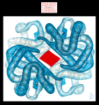
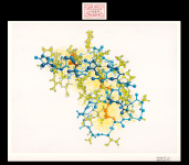
Ribonuclease S
Lysozyme (512)
Crambin
DPG-Hemoglobin Complex
Trypsin
Illustrations are free for use under a CC-BY-4.0 license
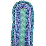
Mycoplasma mycoides

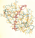
RecA and DNA

Vascular Endothelial Growth Factor (VegF) Signaling
Ebola Virus
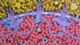
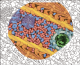
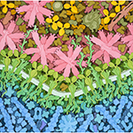
Biosites: Basement Membrane

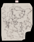
Last Universal Common Ancestor
Influenza Vaccine
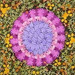
Coronavirus
Excitatory and Inhibitory Synapses

Abiogenesis

Escherichia coli Bacterium
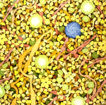
Biosites: Blood Plasma

HIV-Infected Cell

HIV Vaccine
Lipid Droplets

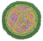
JCVI-syn3A Minimal Cell

Bacteriophage T4 Infection
Autophagy

SARS-CoV-2 and Neutralizing Antibodies

Respiratory Droplet

Myoglobin in a Whale Muscle Cell

Cellulose Synthase
Zika Virus

SARS-CoV-2 Fusion

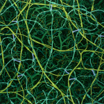
CytoSkeleton

Phage-based COVID-19 Vaccine

SARS-CoV-2 mRNA Vaccine
HIV in Blood Plasma

Caulobacter Polar Microdomain
Poliovirus Neutralization
Immunological Synapse

Red Blood Cell Cytoskeleton
Biosites: Nucleus

Myelin

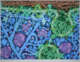
Collagen and Extracellular Matrix
Biosites: Red Blood Cell
Blood
Measles Virus Proteins

Insulin Release

Casein Micelle and Fat Globule in Milk

Insulin Action
Biosites: Muscle
Chloroplast

Model of a Mycoplasma Cell

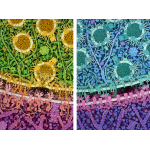
Coronavirus Life Cycle
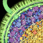
Escherichia coli
Transfer RNA and Gag Protein
Biosites: Cytoplasm
