Videos
Introductions to Protein Structure
![]() | What is a Protein?Proteins play countless roles throughout the biological world, from catalyzing chemical reactions to building the structures of all living things. Despite this wide range of functions all proteins are made out of the same twenty amino acids, but combined in different ways. The way these twenty amino acids are arranged dictates the folding of the protein into its primary, secondary, tertiary, and quaternary structure. Since protein function is based on the ability to recognize and bind to specific molecules, having the correct shape is critical for proteins to do their jobs correctly. Learn more about the relationship between protein structure and function in this video. |
![]() | How Enzymes Work |
![]() | AconitaseAconitase converts citrate into isocitrate as part of the citric acid cycle. |
![]() | Calcium PumpThe calcium pump moves ions across cell membranes allowing the synchronized contraction of muscle cells. |
Structures and Disease
![]() | Penicillin and Antibiotic Resistance
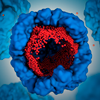
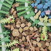
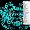
Since its discovery in 1928, penicillin and penicillin-related antibiotics helped save countless lives from bacterial infections. However, in the face of overuse and misuse of antibiotics, bacteria evolved resistance mechanisms that allow them to proliferate even in the presence of the newest antibiotics. |
![]() | Ebola Virus ProteinsTour the molecular anatomy of the Ebola virus with the RCSB PDB. Understanding of the shape and structure of the proteins that make up Ebola is a key component in the fight against the virus. Learn more at Molecule of the Month. |
![]() | A Molecular View of HIV Therapy
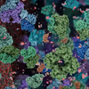
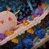
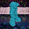
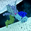
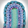
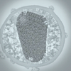
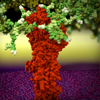
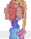
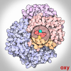
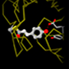
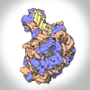
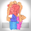
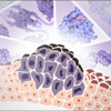
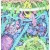
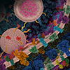
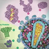
After HIV enters a T-cell, three enzymes play essential roles in the life cycle of the virus. Reverse transcriptase copies the viral RNA genome and makes a DNA copy. Integrase inserts this viral DNA into the cell’s DNA. In the last steps of the viral life cycle, HIV protease cuts HIV proteins into their functional parts. Current antiretroviral drugs target these three enzymes, hindering the virus reproduction. However, enzymes can mutate and become drug resistant, making it vital to use a combination of different drugs that target multiple enzymes. 
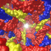
This animation was created using many PDB entries for Reverse Transcriptase (3hvt, 3dlk, 3v6d, 3v4i, 3klg, 3v81), Integrase (3os1, 3os0, 3oya), Protease (3pj6, 1kj4, 1hxb, 2az9, 2azc), HIV Polyprotein (1l6n), Capsid Protein (2m8l), and Matrix Protein (1tam). |
![]() | Learn about HIV from the RCSB Protein Data BankUse the RCSB PDB Resources to Learn about HIV |
![]() | RCSB PDB: An Empowering Resource for Understanding HIV/AIDS at a Molecular LevelThis seminar was presented by Stephen K. Burley, MD, DPhil, and Shuchismita Dutta, PhD on December 5th 2014 as part of the World AIDS Day Symposium at the Rutgers Center for Integrative Proteomics Research in Piscataway, NJ. |
![]() | Staphylococcus aureus and Antibiotic ResistanceHow penicillin and other beta-lactam antibiotics work and what mechanisms the resistant bacteria use it to evade them. |
Animated GIFs
![]() | AconitaseAconitase converts citrate into isocitrate as part of the citric acid cycle. |
![]() | Artificial lattice of a designed DNA crystalDesigned DNA crystal: Small pieces of DNA have been engineered to form a nanoscale lattice. |
![]() | Oxygen Binding in HemoglobinHemoglobin uses a change in shape to increase the efficiency of oxygen transport. |
![]() | Photoactive Yellow Protein and XFEL/SFXStructures of photoactive yellow protein were determined by serial femtosecond crystallography after illumination, capturing the isomerization of the chromophore after it absorbs light. Structures included in this movie include: 5hd3 (ground state), 5hdc (100-400 femtoseconds after illumination), 5hdd (800-1200 femtoseconds), 5hds (3 picoseconds), 4b9o (100 picoseconds), 5hd5 (200 nanoseconds) and 1ts0 (1 millisecond). For more, see the Molecule of the Month on Photoactive Yellow Protein and Guide to Understanding PDB Data: Methods for Determining Atomic Structures |
![]() | Ribosomal SubunitsAtomic structures of the ribosomal subunits reveal a central role for RNA in protein synthesis. Ribosomes are complex molecular machines that build proteins. |
![]() | Vacuolar ATPaseTwo linked molecular motors are used to pump protons across membranes. |
Video Challenge Awards
![]() | 2018 Bacterial Resistance to Beta-lactam AntibioticsFor the fifth year, RCSB PDB invited high school students to tell molecular stories in video. This year's challenge focused on the molecular mechanisms behind bacterial resistance to Beta-lactam antibiotics |
![]() | 2017 DiabetesFor the fourth year, RCSB PDB invited high school students to tell molecular stories in video. This year's challenge focused on the molecular view of the diabetes treatment and management. |
![]() | 2016 DiabetesWinning videos in the 2016 Video Challange for High School Students "Structural Biology & Diabetes" |
![]() | 2015 HIVWinning videos in the 2015 Video Challenge for High School Students "Detecting and Combatting HIV in 3D" |
![]() | 2014 HIVWinning videos in the 2014 Video Challenge for High School Students "Structural Biology of HIV" |
Molecular Animation Guides
![]() | Protein Visualization with Chimera, Blender and AudacityMake your first molecular movie |
![]() | Visualizing PDB Structures with UCSF Chimera for BeginnersLearn the basics of molecular visualization with UCSF Chimera |
![]() | Molecular Animation Q&A |