Creating Paper Models of Protein Domains
Introduction
Proteins are polymers of amino acids. The secondary structure elements are beta sheets, alpha helices and loops. These elements are arranged in 3D space creating the environment for the protein or protein domain to fulfill its function.
As more and more 3D shapes became known, researchers noticed patterns in the folding of whole proteins or protein domains.
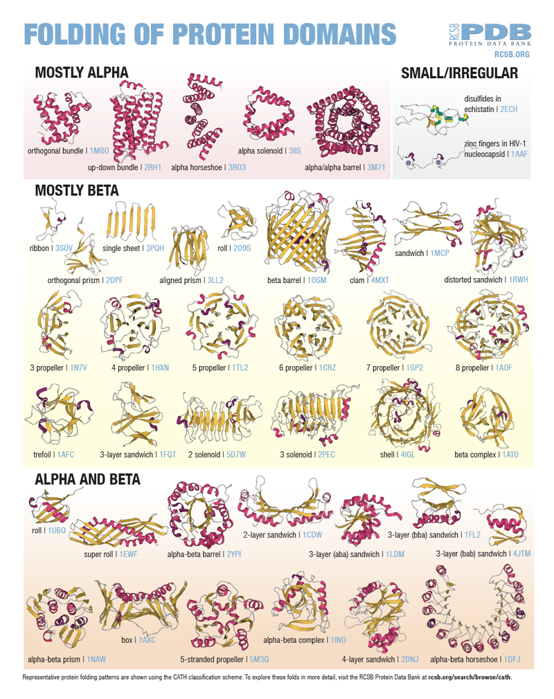
The poster shown here highlights common arrangements of secondary structure elements in protein domains as defined using the CATH classification scheme.
Exploring Common Protein Domains through Building Paper Models
Download printable alpha helices and beta strand templates for hands-on exploration of many protein folding domains.
Using the PDB IDs on the poster, access the corresponding structures on RCSB.org and view in 3D using Mol* to explore and build schematic models of the folds. Use the PDB IDs provided on the poster to explore these domains on RCSB.org in 3D using MolStar (Mol*). Enter the PDB ID in the Import > Download Structure section in the viewer's right menu.
To create an alpha helix, print the helices template as many times as needed (4 pink helices are on each printed sheet). Assemble each helix by cutting on the solid line, folding on the dotted line, tucking the flap inside, and taping it together to form a triangular prism. Each helix comes with an extending loop (gray) that can be used to connect to other elements.
To create a beta strand, print out the template as many times as needed (9 yellow beta strands are on each printed sheet). Cut each strand on the solid line. Each beta strand comes with an extending loop (gray). Multiple strands can be used to form models of beta sheets.

|

|

|
Alpha helix and beta strand prepared from the template |
Building Common Protein Folding Domains
Model 1: TIM Barrel/Alpha-Beta Barrel


The fold is named after the enzyme triose-phosphate isomerase. The fold is also referred to as the alpha-beta barrel. This fold is assumed by about 10% of enzymes.
In this type of fold, the beta strands form a beta barrel in the center, surrounded by alpha helices. The catalytic site of the enzyme is in the middle of the barrel.
Explore the PDB structure 8TIM in 3D to see an example of this fold.
Model 2: Beta Sandwich/Nanobody


This fold features two layers of beta sheets.
Many copies of this fold are connected together with flexible linkers to form the distinctive Y-shape of antibodies.
The nanobody in this paper model is similar to a small portion of an antibody.
Explore an example of this domain fold seen in PDB structure 1MCP.Model 3: ABA Sandwich/Rossman Fold


The Rossman fold is a type of alpha-beta-alpha (ABA) sandwich.
In this fold, 6 parallel beta strands form an extended beta sheet. Two of the alpha helices are under, and two are over the beta sheet.
The fold is common and found in many enzymes that bind to nucleotide cofactors.
Explore an example of this domain fold seen in PDB structure 1LDM.
Learn more
Explore all paper models at PDB-101
