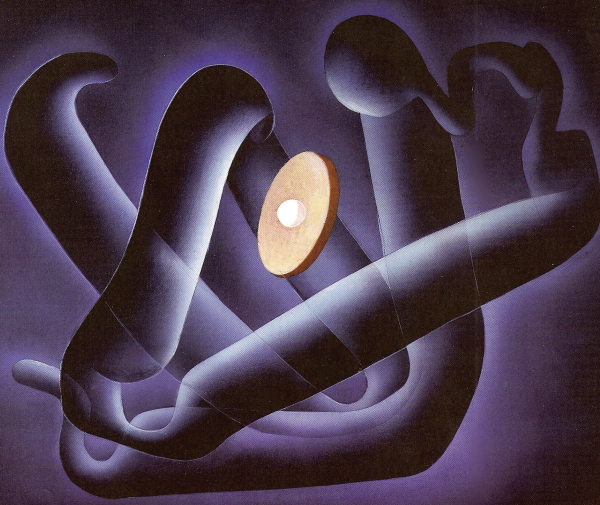
Myoglobin Fold
1987, Unknown Dimensions

Geis illustrated the structure of myoglobin, focusing on the folding pattern of the secondary structure of the protein. Unlike previous myoglobin Illustrations, this painting focuses on the tertiary structure of the molecule rather than the sequence or surface.
Used with permission from the Howard Hughes Medical Institute (www.hhmi.org). All rights reserved.

Related PDB Entry: 1MBN
Experimental Structure Citation
Kendrew, J., Bodo, G., Dintzis, H., Parrish, R., Wyckoff, H., & Phillips, D. (1958). A Three-Dimensional Model of the Myoglobin Molecule Obtained by X-Ray Analysis. Nature, 181, 662-666. DOI:10.2210/pdb1mb/pdb
About Myoglobin
Molecule of the Month: Myoglobin
Myoglobin stores oxygen in cells, and is particularly plentiful in muscle cells. This historic structure (sperm whale myoglobin) was the first structure of a protein, solved in the laboratory of John Kendrew.
Text References
Goodsell, D. (2000). Molecule of the Month: Myoglobin. DOI: 10.2210/rcsb_pdb/mom_2000_1


