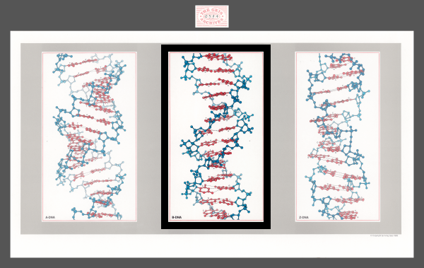
B-DNA
1983
Geis’ depicted version of the B-helix is the most common form of DNA. The major and minor grooves of the helix are shown clearly.


Used with permission from the Howard Hughes Medical Institute (www.hhmi.org). All rights reserved.

Related PDB Entry: 1BNA
Experimental Structure Citation
Drew, H. R., Wing, R. M., Takano, T., Broka, C., Tanaka, S., Itakura, K., & Dickerson, R. E. (1981). Structure of a B-DNA dodecamer: conformation and dynamics. Proc. Natl. Acad. Sci. USA, 78, 2179-2183.
About B-DNA
The DNA B-helix is the most common form of DNA. It exists under neutral pH and physiological salt conditions ("B-Form, A-Form, Z-Form of DNA" website). It is right-handed and consists of 10 base pairs per turn.
Note: The depicted Chimera and Jmol structures (consisting of 12 base pairs) are predictions by Dr. Wilma Olsen based on the crystallized fragment (consisting of 4 base pairs) to match Geis’ illustration (Zheng et al., 2009).
Text References
B-Form, A-Form, Z-Form of DNA. University of California, Davis. URL: http://biowiki.ucdavis.edu/Core/Genetics/Unit_I%3A_Genes%2C_Nucleic_Acids%2C_Genomes_and_Chromosomes/2%3A_Structures_of_nucleic_acids/B-Form%2C_A-Form%2C_Z-Form_of_DNA
Zheng G., Lu X. J., & Olson W. K. (2009). Web 3DNA—a web server for the analysis, reconstruction, and visualization of three-dimensional nucleic-acid structures. Nuc. Acids Res. 37 (Web Server Issue), W240-W246
