Molecular Landscapes by David S. Goodsell
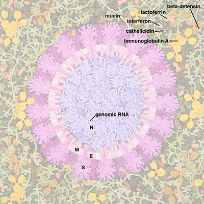
Coronavirus, 2020

Acknowledgement: Illustration by David S. Goodsell, RCSB Protein Data Bank; doi: 10.2210/rcsb_pdb/goodsell-gallery-019
This painting depicts a coronavirus just entering the lungs, surrounded by mucus secreted by respiratory cells, secreted antibodies, and several small immune systems proteins. The virus is enclosed by a membrane that includes the S (spike) protein, which will mediate attachment and entry into cells, M (membrane) protein, which is involved in organization of the nucleoprotein inside, and E (envelope) protein, which is a membrane channel involved in budding of the virus and may be incorporated into the virion during that process. The nucleoprotein inside includes many copies of the N (nucleocapsid) protein bound to the genomic RNA.
For more information about coronavirus, visit the February Molecule of the Month on coronavirus proteases. There are also many structures of coronavirus proteins in the PDB archive; search for "coronavirus" at the main PDB site to see them.
The painting is based on information about the SARS virus, taken primarily from the following publications:
Masters PS (2019) Coronavirus genomic RNA packaging. Virology 537, 198-207.
Surya W, Li Y, Torres J (2018) Structural model of the SARS coronavirus E channel in LMPG micelles. BBA Biomembranes 1860, 1309-1317.
Li F (2016) Structure, function, and evolution of coronavirus spike proteins. Annu. Rev. Virol. 3, 237-261.
Chang CK, Hou MH, Chang CF, Hsiao CD, Huang TH (2014) The SARS coronavirus nucleocapsid protein - forms and functions. Antiviral Res. 103, 39-50.
Neuman BW, Adair BD, Yoshioka C, Quispe JD, Orca G, Kuhn P, Milligan RA, Yeager M, Buchneier MJ (2006) Supramolecular architecture of severe acute respiratory syndrome coronavirus revealed by electron cryomicroscopy. J. Virol. 80, 7918-7928.
More information on the painting is available in an article at PLoS Biology.
