Bacteriophage T4 Infection
JCVI-syn3A Minimal Cell
Insulin Release
HIV Vaccine
Caulobacter Polar Microdomain
HIV-Infected Cell
Escherichia coli Bacterium
Cellulose Synthase
Myoglobin in a Whale Muscle Cell
Collagen and Extracellular Matrix
CytoSkeleton
Transfer RNA and Gag Protein
RecA and DNA
Casein Micelle and Fat Globule in Milk
Model of a Mycoplasma Cell
Phage-based COVID-19 Vaccine
Coronavirus
Immunological Synapse
Coronavirus Life Cycle
Myelin
Respiratory Droplet
SARS-CoV-2 and Neutralizing Antibodies
SARS-CoV-2 Fusion
SARS-CoV-2 mRNA Vaccine
Red Blood Cell Cytoskeleton
Measles Virus Proteins
Lipid Droplets
Poliovirus Neutralization
Influenza Vaccine
Excitatory and Inhibitory Synapses
Abiogenesis
Last Universal Common Ancestor
Zika Virus
Insulin Action
Ebola Virus
Autophagy
Mycoplasma mycoides
Chloroplast
Vascular Endothelial Growth Factor (VegF) Signaling
Biosites: Basement Membrane
Biosites: Red Blood Cell
Biosites: Blood Plasma
Biosites: Cytoplasm
Biosites: Muscle
Biosites: Nucleus
Blood
HIV in Blood Plasma
Escherichia coli

Molecular Landscapes by David S. Goodsell

Blood, 2000

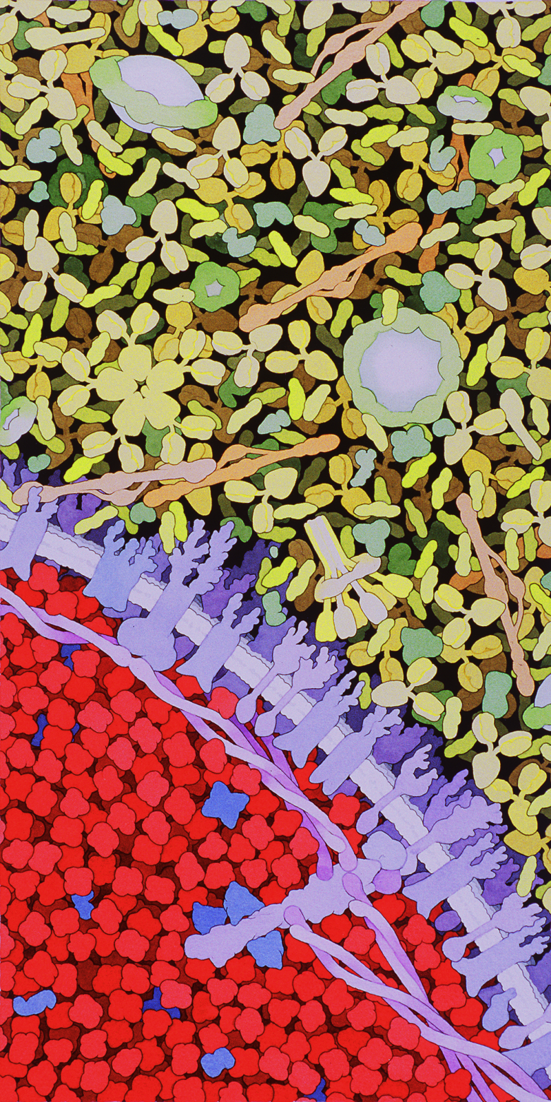
Acknowledgement: Illustration by David S. Goodsell, The Scripps Research Institute. doi: 10.2210/rcsb_pdb/goodsell-gallery-003

This illustration shows a cross-section through the blood, with blood serum in the upper half and a red blood cell in the lower half. In the serum, look for Y-shaped antibodies, long thin fibrinogen molecules (in light red) and many small albumin proteins. The large UFO-shaped objects are low density lipoprotein and the six-armed protein is complement C1. The red blood cell is filled with hemoglobin, in red. The cell membrane, in purple, is braced on the inner surface by long spectrin chains connected at one end to a small segment of actin filament.

beta